Large-Area High-Resolution Imaging
Highest-resolution, accurate SEM imaging of large samples exceeding a single typical field of view (in the order of a few tens of μm) is a challenging procedure. A set of several hundreds or thousands of images have to be stitched together in order to display an area spanning several millimeters, or even centimeters, as a “Google Earth”-style map. Any standard mechanical stage, even followed by software optimization, will produce visible stitching and invisible placement errors and thus yield distorted image mosaics.
Raith systems are different: By reversing the functionality of a professional electron beam lithography system, the sample surface is not exposed; instead, existing nanostructures are seamlessly imaged using the extreme placement accuracy of the systems stage and electron beam.
Users can benefit e.g. from the “on-board” Laser Interferometer Controlled Stage technology, related field of view alignment functionality, and drift correction algorithms. These features deliver ultra-precise and fully automated image acquisition for generating highly accurate and undistorted “land maps” of large samples with highest resolution and stitching accuracy in the nm range in 2D – or 3D in case multiple (sequentially deprocessed) layers are involved.
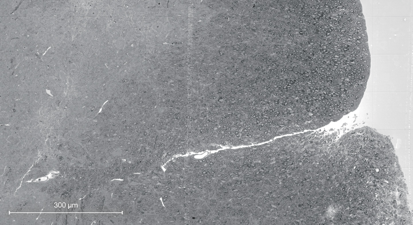
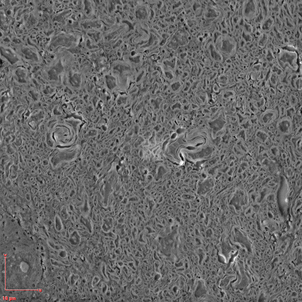
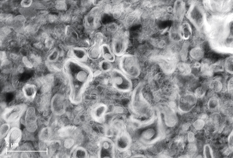
Reverse Engineering and Connectomics
Chip manufacturers are not the only users to rely on Raith technology for reverse engineering applications and identification of counterfeiting or parasitic chips. Nanobiologists, too, can use all these features to reveal the brain circuitry in connectomics. Here, analysis of the connectivity of neurons is the driver for (3D) brain mapping. CHIPSCANNER helps to understand the complex architecture of a spinal cord by providing a high-resolution mosaic of the biological sample.
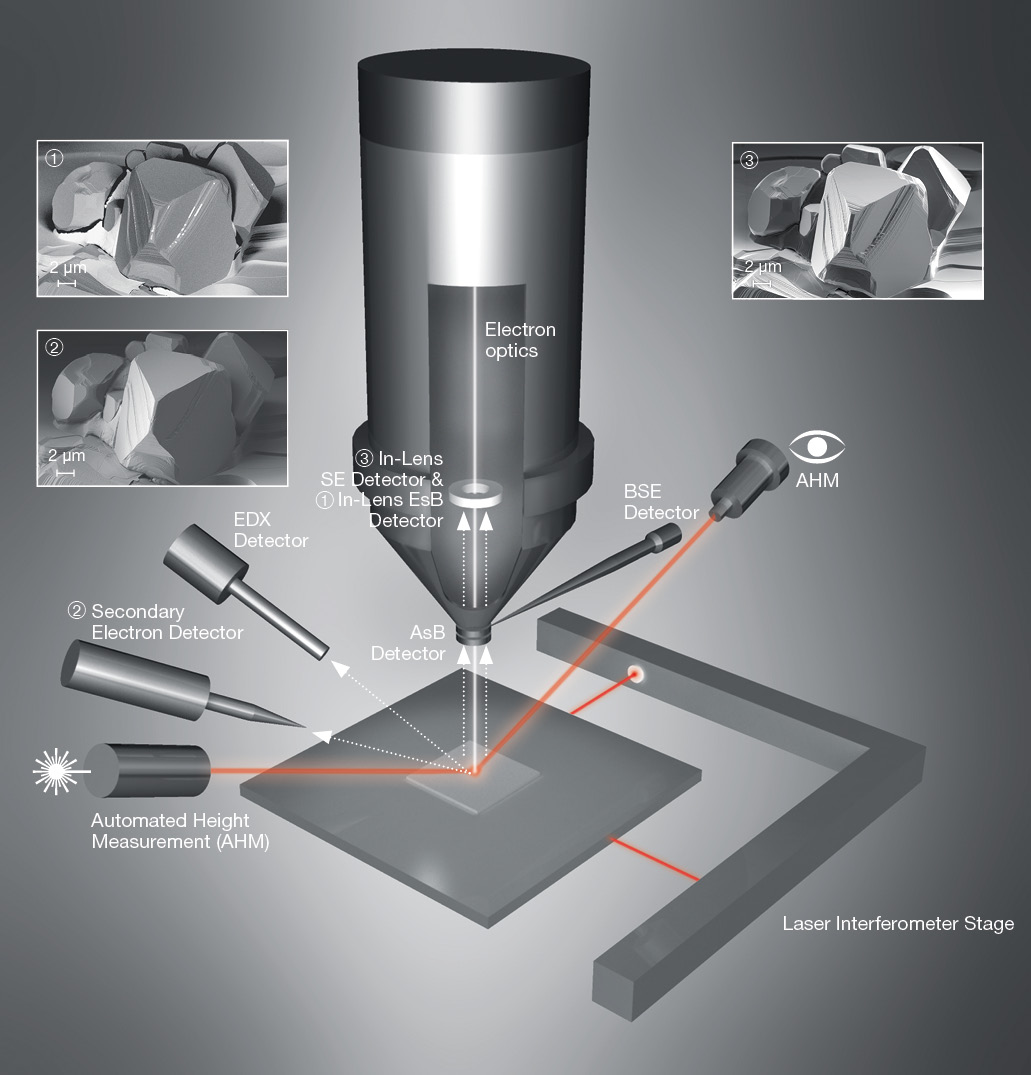
True Large-Area SEM with various detectors
The Laser Interferometer Stage is undeniably an important ingredient of any professional nanolithography system. In reversing the functionality of an EBL tool from writing to imaging, it is also a critical component for accurately stitching SEM images over large areas because it allows the sample to be accurately positioned under the field of view (FOV). Moreover, it is used to map out errors in the FOV to a placement accuracy of <1 nm.
Various detectors – whether secondary or backscattered detectors – help to achieve the best image contrast or extract the optimum analytical information. Laser height measurement and corresponding automatic sample height adjustment to an accuracy of better than 1 μm ensures focus stability over an entire sample (depending on the topography) such as a large wafer, even if the sample is inclined or exhibits a strong curvature such as a wafer bow.