Introduction
IONMASTER is a novel ion microscope with a dedicated magnetic sector Secondary Ion Mass Spectrometer (SIMS). It is the perfect tool for the highest resolution SIMS imaging, offering unparalleled sensitivity in detecting even the smallest features. Experience a revolution in nanotechnology, opening new frontiers for a wide range of applications.
Benefits
Experience FIB-SIMS beyond limits
Highest spatial resolution and sensitivity
IONMASTER offers various application-specific primary ions to optimize results. Dedicated reactive primary ions enable the best ionization yield, and light or heavy ions adjust the sputter yield. IONMASTER magSIMS, with its sophisticated extraction and transfer optics, ensures the highest transmission of secondary ions, guaranteeing optimal performance for any analytical challenge.
Shortest time to result
Featuring a focal plane detector that acquires full mass spectra of each pixel simultaneously and a magnetic sector for mass separation without duty cycles, it delivers results in the shortest time. In combination with a vast selection of primary ions, the sputter yield and ionization of secondary ions can be optimized. Use light ions to minimize sample damage while maintaining the highest lateral resolution, or choose heavy ions for the highest depth resolution and high sputter yield.
SIMS analysis with ion microscopy and nanofabrication
Correlative SIMS and SE ion imaging for multimodal analysis enables comprehensive sample information. The high-precision laser interferometer stage allows for precise CAD-based multisite analysis and blind navigation for the analysis of pristine sample areas. Advanced nanofabrication workflows and milling strategies ensure superior analytical performance, elevating ion nanofabrication capabilities to the next level.

Ultra
Positioning

Mag
SIMS

Ion
Select
Software
A new era in nano-characterization and imaging

User interface for SIMS analysis
IONMASTER OPS software controls all features and hardware components of the SIMS spectrometer.
IONMASTER VIEW software is used for rapidly viewing the acquired data files and performing measurements and data processing.
Furthermore, the proprietary Digital RAITH Nanosuite software operates all nanofabrication and imaging system functionalities.
IONMASTER VIEW software is used for rapidly viewing the acquired data files and performing measurements and data processing.
Furthermore, the proprietary Digital RAITH Nanosuite software operates all nanofabrication and imaging system functionalities.
Positioning and adjustment of the extraction system
Automated movement of transfer optics controls all voltages of SI optics. Additionally, switching between positive and negative SIMS analysis modes can be done within a few seconds.
Data acquisition for different analysis modes
Various analysis modes available: Live mode, Total Ion Count (TIC) imaging, Focal Plane Detector (FPD) mass spectrum recording, FPD depth profiling, FPD imaging, and FPD 3D imaging.
Magnet control
The magnetic field in the magnetic sector can be adjusted to select the desired mass range of the detected secondary ion.
Focal plane detectors
Simple and quick adjustment of the parameters to optimize the data acquisition of full continuous spectra.
100 person-years of software programming
Technical data
Lateral SIMS imaging resolution
< 20 nm
Dynamic range / Sensitivity
107 / < 10 ppm
Polarity of secondary ions
selectable,
positive / negative
positive / negative
SE Imaging resolution by using the TIC in the negative mode
≤ 5 nm
Detectable elements / isotopes
All
SIMS depth resolution
< 10 nm
Are you interested in more details and insights?
IONMASTER brochure (.pdf)
Application use cases
Highest-resolution SIMS imaging and depth profiling
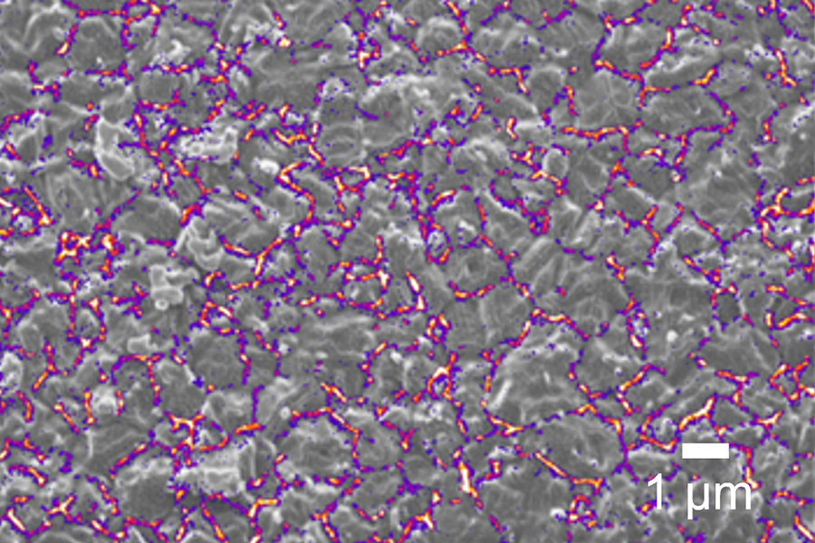
Multimodal analysis: Ion induced SE surface signal (gray) of a Rubidium (Rb) treated CIGS solar cell with Rb SIMS overlay (purple)
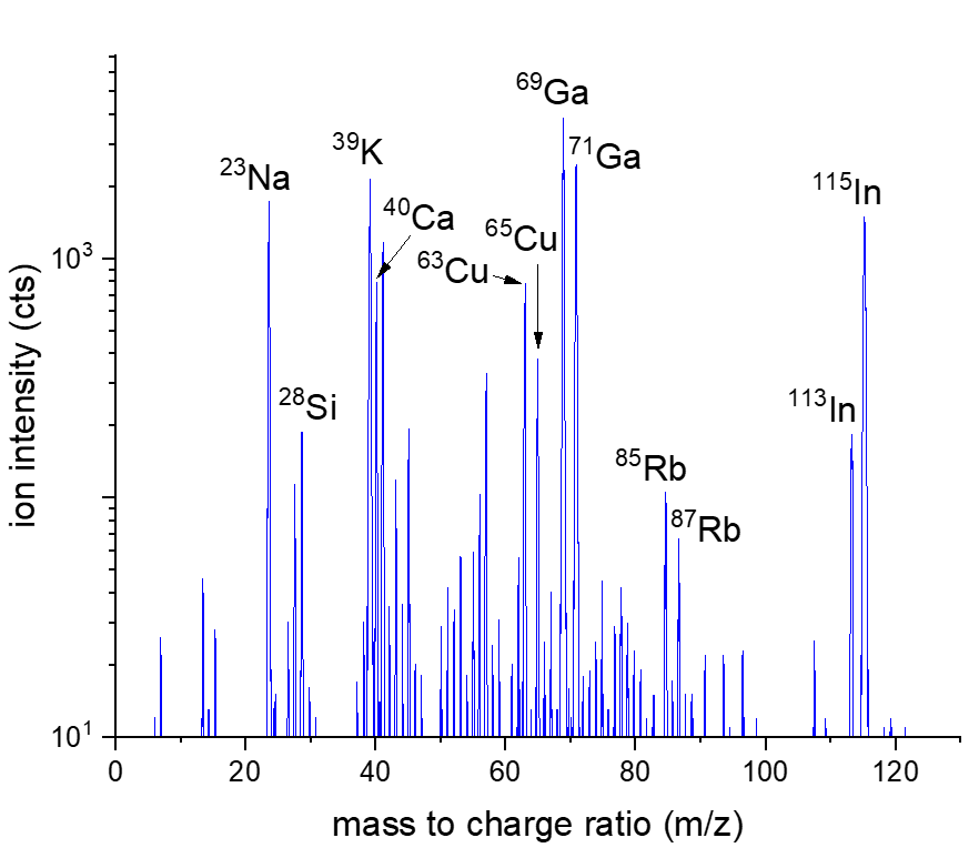
Mass spectrum of CIGS solar cell in positive mode. A full mass spectrum is acquired for each pixel during SIMS mapping
3D SIMS reconstruction of the Rubidium(85Rb & 87Rb) isotopes (orange) and Indium(113In & 115In) (gray) isotopes. Rubidium is segregated in the grain boundaries of the CIGS surface

Mouse gut cells with nanoparticles: Aluminum nanoparticles (red) are clearly detected and resolved outside the cells, showing distribution around the cell body
3D SIMS reconstruction of Ni (red) – Ti (green) multilayer micropillars on Cr substrate (blue), diameter 1um, layer thickness 10 nm – 50 nm
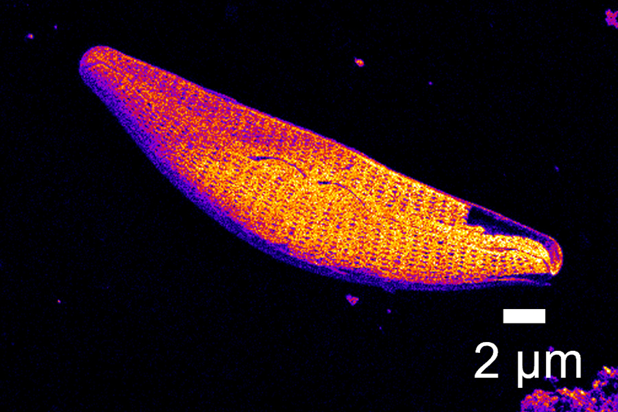
SIMS image of a diatom composed of Si and SiO2 signal multiplexing

Ion images revealing the high depth of field and high image resolution. Li ion beam image of Bi2 Ca2Co compound material

Li ion image of diatoms
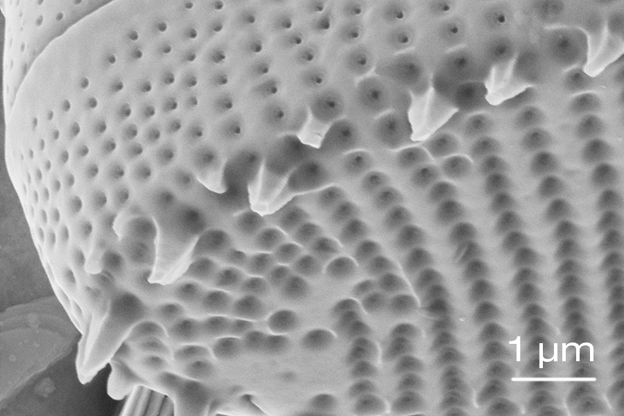
Si ion image of diatoms
Solutions
RAITH offers integrated solutions that increase performance and create market opportunities. In any production environment.

Mix and Match
Seamless blending of laser direct writing, ion beam, and electron beam technologies, unlocking new nanostructuring horizons.