Introduction
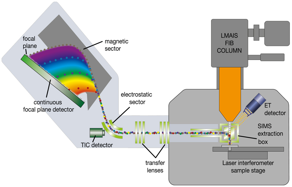
The MagSIMS consists of a dedicated secondary ion extraction optics followed by post-acceleration optics, transfer optics, a modified Mattauch-Herzog double focusing magnetic sector mass analyzer and a continuous focal plane detector for parallel detection of all ions.
It is designed and integrated in such a way that the secondary ion extraction optics can be inserted in between the sample and the objective lens of the FIB column during SIMS operation, while being retracted to a parking position on the side of the chamber for normal FIB operation.
It is designed and integrated in such a way that the secondary ion extraction optics can be inserted in between the sample and the objective lens of the FIB column during SIMS operation, while being retracted to a parking position on the side of the chamber for normal FIB operation.
Experience unparalleled visualization of nanoscale structures and chemical information
Key features
For highest sensitivity and transmission.
Records a full mass spectrum of each pixel. Easy and fast alignment of detection system, excellent data post-processing capabilities.
No duty cycles, very short data acquisition times.
Benefits for applications
MagSIMS takes nano characterization to the next level
Fast time to result
Continuous detection with magnetic sector, high signal / sensitivity through extraction and transfer optics.
Highest resolution and sensitivity
MagSIMS with its sophisticated extraction and transfer optic ensures highest transmission of secondary ions, guaranteeing optimal performance for any analytical challenge.
Ease of use
Easy and fast setup of SIMS analysis.
Technical details
Precision technology developed to challenge the frontiers of nanotechnology
Mass spectrometer
MagSIMS features a double focussing Mattauch-Herzog mass spectrometer for the precise measurement of the mass-to-charge ratio (m/z) of ions. The advantage of a double-focusing mass spectrometer is high-resolution capability and accurate measurements of m/z ratios for a wide range of ions. This makes it particularly useful for applications in analytical chemistry, isotope ratio measurements, and the study of complex mixtures of compounds.
Results
Detection of all elements and isotopes
Easy detection of light elements like Hydrogen or Lithium
Reliable identification of nanoparticles
Analysis of thin interfaces and films
3D chemical analysis at the nanoscale
Application use cases
3D SIMS reconstruction of the Rubidium (85Rb & 87Rb) isotopes (orange) and Indium (113In & 115In) (gray) isotopes. Rubidium is segregated in the grain boundaries of the CIGS surface
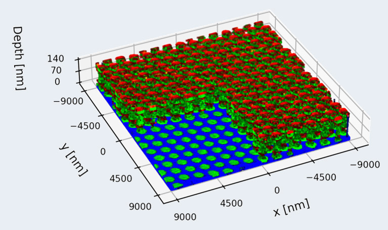
3D SIMS reconstruction of Ni (red) and Ti (green) multilayer micropillars on a Cr substrate. Diameter 1 μm, layer hickness 10 nm – 50 nm

Corresponding depth profile of the micropillars

SIMS images of 27Al and 51V distribution of a biomedical implant obtained in positive SIMS mode. Material: Ti-6AI-4V alloy

Hydrogen distribution of the same biomedical implant acquired in negative SIMS mode.
Sample courtesy of Athira Suresh Kumar (Luxembourg Institute of Science and Technology)
Sample courtesy of Athira Suresh Kumar (Luxembourg Institute of Science and Technology)

Mouse gut cells with nanoparticles. Aluminium nanoparticles (red) are clearly detected and resolved outside the cells showing the distribution around the cell body.
